


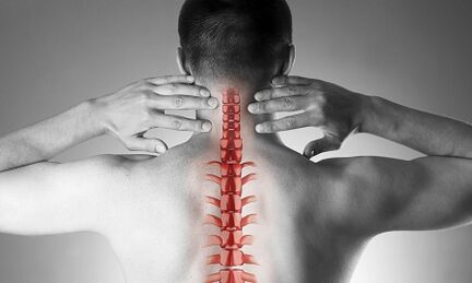
Cervical osteochondrosis or spondylosis occurs as a result of changes in the form and structure of the vertebrae.Despite the fact that the cervical region is short enough in connection with the total length of the spine, it is probably the most important part of the spine.Each pair of adjacent vertebrae forms the intervertebral holes through which the nerve roots go and go to every muscle and organ of the upper half of the body.Through other holes - in the lateral processes of these vertebrae - vital vessels guarantee the blood supply to the brain.
The causes of osteochondrosis of the cervical spine
The causes of osteochondrosis are:
- Injuries,
- "Stuck" work on the monitor located below eye level,
- physical labor associated with the transfer of weights,
- Long -term staying car driving,
- Work by phone without using distance devices (in this case the operator presses the phone to the ear arm)
- Constitutional characteristics (curves, congenital changes in the cervical vertebrae, short neck)
Formation of pathological vertebrae changes
With osteochondrosis, small spots begin to form at the edges of the vertebrae bodies, which can injure the structures located nearby.Most often, this happens in response to the excess load of the cervical ward and is not only the result of the "aging" of the intervertebral joints (recall that it was considered degenerative osteochondrosis, then a natural "age -related age", such as osteaterrosis).With the development of the disease, the plates to close the vertebrae appear and reduce the height of the intervertebral discs.These discs are normal, play the role of the shock absorber between the vertebrae and, among other things, prevent damage to the spinal roots.With progressive osteochondrosis, a convex (hernia) of the jacket is strong on the intervertebral disc, on which there is more and more pressure during the disease, while weakening the "restriction" of connections on all sides.This hernia is also capable of draining the spinal structures and causing neurological manifestations of the disease.
What are the symptoms of cervical osteochondrosis?
Osteochondrosis of the cervical spine with pain syndrome
Any neck pain forces the pathology of the cervical spine.In terms of growth, the intensity of the pain syndrome is divided into 4 stages, the first patient experiences numbness, numbness, a feeling of "tightness" in the area of a particular muscle group, at the fourth stage - the most severe - the pain is so intense that they lead to the immobility of the patient and the loss of effectiveness.
In addition to the cervical and dorsal pain syndrome, the patient notes "reflected" (radiating) pain in the upper limb, sub -islets lateral areas of the breast.
Osteochondrosis of the cervical spine with radicular syndrome
They speak of participation in the process of nerve roots when pain, numbness and numbness spread to the lower jaw, upper back, forearm and fingers.At the same time, the patient draws attention to the fact that "as if he was leaving," he was sleeping uncomfortable.The morning stiffness in the joints of the fingers, which lasted no more than 10-15 minutes, is noted.With the development of radicular syndromes, a decrease in the muscular strength of the upper limbs may be noted during examination.
Osteochondrosis of the cervical spine with "spinal artery syndrome"
Regarding involvement in the process of blood vessels (pressing them with hernial convexity or osteophyte), they say that when the patient complains of frequent headache attacks, especially after a long stay in a certain position when he is thrown out of his head (for example, when you swim with flour), if worn in ears, they are worn.This clinical situation is well detected with the help of ultrasound (with "Doppler mapping mode").Ultrasound determines the inquisition of the spinal arteries, narrowing their lumen.In this case, we can talk about surgery, since the pronounced change in the bloodstream in the vertebrae is a risk factor for the development of stroke.
Osteochondrosis of the cervical spine with "heart (heart) syndrome"
This syndrome forces the patient to contact the cardiologist mainly, as the main complaints are related to the pain in the left half of the breast, the sub -kapular area, which weakens or intensifies when physical activity or body position is performed.After the exclusion of myocardial infarction and other heart disease, the patient falls under the supervision and treatment of a neurologist and orthopedist.
Diagnostics
To clarify the diagnosis, four methods are used: radiography, ultrasound, computed tomography and magnetic resonance imaging.
The most affordable method is still radiography of the cervical spine, the most information is radiography in lateral projection ("side view").This method allows for the first approximation to determine the presence of injury, gross structural changes in the vertebrae.
Ultrasound (ultrasound) is performed to clarify the condition of the vertebrae arteries.With the help of this method, they discover whether blood flow is disturbed and, if so, to what extent and what type of obstacle they occur and where they are localized.
Computed tomography (CT).It allows you to evaluate the condition of the bone structures, the degree of bone density, allows you to see smaller osteophytes (bone growths) than possible in x -ray.
Magnetic resonance imaging (MRI).This type of examination is indispensable for suspected hernia, accurate localization of spinal cord damage and the degree of this damage.This study is necessary if the issue is raised by surgical (surgical) treatment of diseases of the cervical spine.
Treatment of cervical osteochondrosis
Medication
The standard set of products for the treatment of cervical osteochondrosis reflects the purpose of treatment: pain relief syndrome, elimination of painful muscle spasm and inflammation of the nerve roots, increasing the mobility of the spine.In order to achieve these goals, mainly the use of painkillers, NSAIDs -non -steroidal anti -inflammatory drugs, muscle relaxants are used.It should be remembered that the self -replacement of these groups can be dangerous as there is a possibility of misinterpreting the symptoms as well as an underestimation of the side effects of these drugs.Local (Basel) Medicines among NSAIDs in the form of gels are widely used and if the pain is stopped, the same medicines can now be used in the form of ointments.
Systemic drugs are used for the treatment of osteochondrosis at a deeper, "basic" level.These substances restore the cartilage structures of the vertebrae, prevent their more damage.The treatment courses are long, the effect lasts for many months.
Cervical osteochondrosis has significant differences from the pathology of the other spine.Neck pain in this case may not be triggered by signals from the spinal nerves suffering, but by a painful chronic muscle over -tension - all together are called muscle syndrome.This is a completely "benign" condition that is well treated with the same set of medicines: non -steroidal anti -inflammatory drugs, muscle relaxants, using intramuscular "blockage" using steroids.Usually, the doctor reveals acute pain when examining so -called "triggering" points throughout the cervical spine, as well as in the muscles of the upper shoulder.More often, such pathology is found in women, mostly younger than 40 years.Despite the pronounced pain syndrome, the structures of the vascular nor remain intact, the blood flow to the area of the head does not suffer.
Manual therapy
This method of treatment may be effective for recently occurring (often as a result of a small injury, subluxation) neck pain that is not accompanied by dizziness, other changes in the nervous system and the circulatory system.It is permissible to resort to manual therapy only after a thorough examination, in addition, the doctor performing this procedure should have sufficient experience in the field of traumatology and orthopedics.With "old" forms of the disease, the use of manual therapy is dangerous!
Two methods of this type of intervention are known:
- manipulation (sharply short influences of significant force aimed at eliminating subluxation, well -known "bone clicks");
- Mobilization (the method is based on a smooth stretch stretch after warming and relaxing of the muscle corset of the neck).
A combined method is also used based on a combination of two main ones.It is important to remember that in addition to these contraindications, manual therapy is prohibited for any disease, accompanied by an increase in blood pressure, for any pathology of the thyroid gland and Ent or ent -ord.
Treatment of cervical osteochondrosis at home
Medical gymnastics for cervical osteochondrosis
The first and basic rule for beginners to do physiotherapy exercises is not to perform exercises, overcoming painful sensations.Of course, you should not start in the "acute" period when the pain has just appeared.Another important recommendation is to avoid sudden movements and circular movements in the cervical region.
Each lesson should start with a short light for self -government of the neck muscles.
The following is a "warming" warm -up:
- The arms are lowered to the body, the shoulders are uniform, the back is straight (you can check the posture with heels, shoulder blades and an ass to the wall).We walk in the place for 1 min on the whole leg, 1 min - on socks, 1 min - on the heels.
- The starting position is the same.We squeeze the brushes in our fists, raising our shoulders, our hands upright.The movements are slow, we do 20 reps, the last elevation is more than 5 seconds.We make sure that the neck muscles are not "fixed".
- The starting position is the same.We tilt our heads in turn to the right, then on the left.The movements are smooth, one slope of 8 accounts, at the end point of slope - hold for 8 seconds.
- The starting position is the same or sits in a hard chair.Smooth head slopes forward, at the end point - hold for 8 seconds
- The starting position is the same or sits in a hard chair.Slowly tilt your head forward while the chin in the chest, then slowly rotate the head to the right (to 4 accounts) and left (to 4 accounts).Do not allow muscle tension.
- The starting position is the same or sits in a hard chair.We lift your shoulders up to 4 accounts, and then lower them into 4 pieces.10 reps.
- The starting position is the same or sits in a hard chair.We lift our shoulders, but now we perform circular motions in the front, 8 accounts.10 reps.
- We align the back, check the stand.Up to 4 accounts, we reduce the shoulder blades behind your back, trying to connect them, at the end point where we stay for 8 seconds, then return to the starting position.
Pillows
As mentioned earlier, the hypertension of the neck muscles is the first and often the main cause of the development of cervical osteochondrosis.The rational selection of pillows and mattresses, providing a quiet and comfortable position during sleep is no less than gymnastics, physiological activity and drugs.
When choosing a mattress, pay attention to the composition of the filler (the products are appropriate, at least half of the coconut chip, that is, there is a sufficient degree of hardness).Soft spring mattresses do not provide sufficient straightening of the spine.The most optimal sleep for sleep is on the side, pull one or both knees to the stomach.The pillow should be located in such a way as to fill the entire space between the shoulder, the ear and the mattrates, the parietal part (the crown) of the head is on the same horizontal line with the spine.To avoid too high and too low as well as soft pillows.The ideal option is an ergonomic product, that is, in this case, with a small squeezing pound on one side.
General recommendations
Pay attention to the stand.During walking or upright, the position is a position when the breasts protrude forward and the stomach is pulled.
Avoid the long -term sitting in a sitting position.A simple rule is known to prevent cervical osteochondrosis: after every 60 minutes, 10-15 minutes of walking or heating is required.
The chairman for work must have a high back or back.
In a sitting position, the legs should rest on the floor and the neck should not be tense.For this purpose, use special orthopedic devices: rollers under your neck when driving in a car under your back.
Avoid lifting weight.If necessary, the knees down, press the heavy object to the body and then stood smoothly, using the strength of the muscles of the legs, but not the "thrust" on the back.
Do not bend with your feet upright.Use stands, work surfaces to bring the object closer to yourself, not to persuade your face to the subject.Try to do homework sitting on a chair or gymnastic ball.
If you need to use a mop, broom or rake, do not strain your arms, back and neck, do not bend away.
Avoid brass -style swimming.